MRI- Magnetic Resonance Imaging
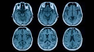
Magnetic resonance imaging (MRI) is a medical imaging procedure that uses strong magnetic fields and radio waves to produce cross-sectional images of organs and internal structures in the body. Because the signal detected by an MRI machine varies depending on the water content and local magnetic properties of a particular area of the body, different tissues or substances can be distinguished from one another in the study image.
MRI can give different information about structures in the body than can be obtained using a standard x-ray, ultrasound, or computed tomography (CT) exam. For example, an MRI exam of a joint can provide detailed images of ligaments and cartilage, which are not visible using other study types. In some cases, a magnetically active material (called a contrast agent) is used to show internal structures or abnormalities more clearly, resulting in high contrast MR images. In most MRI devices, an electric current is passed through coiled wires to create a temporary magnetic field around a patient’s body. (In open-MRI devices, permanent magnets are used.) Radio waves are sent from and received by a transmitter/receiver in the machine, and these signals are used to produce digital images of the area of interest.
Usage Many diseases, such as certain brain tumors, can be visualized using MRI because of a high contrast definition, which does not always require contrast agents to produce detailed images of blood vessels. MRI scanners can image a wide range of body parts including injuries of the joints, the blood vessels, the breast, as well as abdominal and pelvic organs such as the liver or reproductive organs. Using MRI scans, physicians can diagnose or monitor treatments for a variety of medical conditions, including:
- Abnormalities of the brain and spinal cord
- Tumors, cysts, and other abnormalities in various parts of the body
- Injuries or abnormalities of the joints
- Certain types of heart problems
- Diseases of the liver and other abdominal organs
- Causes of pelvic pain in women (e.g. fibroids, endometriosis)
- Suspected uterine abnormalities in women undergoing evaluation for infertility
Risks/Benefits MRI does not use ionizing radiation (high-energy radiation that can potentially cause damage to DNA, like the x-rays used CT scans). There are no known harmful side-effects associated with temporary exposure to the strong magnetic field used by MRI scanners. However, there are important safety concerns to consider before performing or undergoing an MRI scan:
- The magnet may cause pacemakers, artificial limbs, and other implanted medical devices that contain metal to malfunction or heat up during the exam.
- Any loose metal object may cause damage or injury if it gets pulled toward the magnet, because it can fly at a speed of 35-40 miles/h.
- If a contrast agent is used, there is a slight risk of an allergic reaction. MRI contrast agents can cause problems in patients with significant kidney disease.
- Dyes from tattoos or tattooed eyeliner can cause skin or eye irritation.
- Medication patches can cause a skin burn.
- The wire leads used to monitor an electrocardiogram (ECG) trace or respiration during a scan must be placed carefully to avoid causing a skin burn.
- Prolonged exposure to radio waves during the scan could lead to slight warming of the body.
US-FDA